dc74a9ce5b9ab55fdd1d268a40d2ba10
| License | http://creativecommons.org/licenses/publicdomain/ |
| Rating | 2.5 |
| Vetted | Trusted |
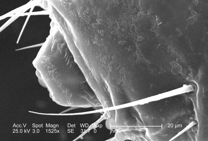
| Description | Magnified 1525x, this 2006 scanning electron micrograph (SEM) depicted an enlarged dorsal view of the mouth region of a male louse, Pediculus humanus var. corporis. In this particular view, the louses haustellum is withdrawn, or internalized inside the mouth region of the insects cephalic region. The haustellum is a tube-like structure, fromed from what is believed to be a modification of the labium, but is armed with teeth, which act to grab, and hold on to the skin surface of its host while obtaining its blood meal. The insect pierces the hosts skin with its sharply-pointed set of three stylets, which together for what is termed the fascicle. Note PHIL# 9214, 9216, in which case a lose is in its feeding mode on the skin surface of its host. Created: 2006 |
| Original URL | http://phil.cdc.gov/PHIL_Images/9229/9229_lores.jpg |
| photographer | Janice Carr |
| provider | Public Health Image Library |
